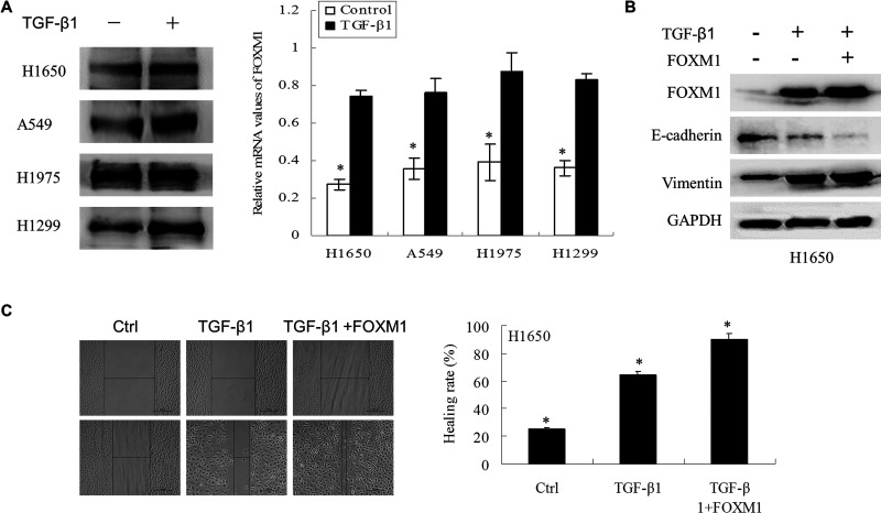
Figure 2.
FOXM1 enhanced TGF-β1-induced EMT in NSCLC cells. (A) NSCLC cells treated with TGF-β1 (5 ng/ml) for 2 days were analyzed by Western blotting and RT-PCR to detect protein and mRNA changes in FOXM1. (B) Vector and FOXM1 stably overexpressing H1650 cells were treated with TGF-β1 (5 ng/ml) for 2 days. The proteins were then extracted, and Western blotting was performed to detect FOXM1, E-cadherin, and vimentin. (C) Wound-healing assays were used to detect the migration capacity of H1650 cells after treatment with TGF-β1 (5 ng/ml) for 3 days in the vector and FOXM1 groups.

