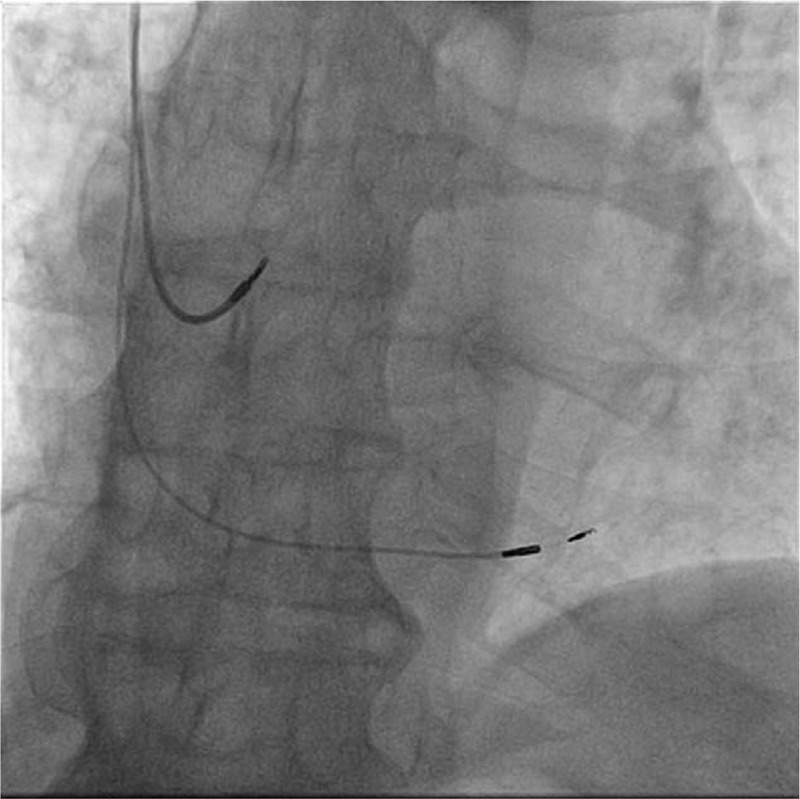
Figure 2.

Fluoroscopic antero-posterior view of the atrial lead and deep interventricular septal lead. Compared to His bundle pacing, in left bundle branch area pacing the active lead is moved 1,5 cm more apically and screwed deep into the interventricular septum.
