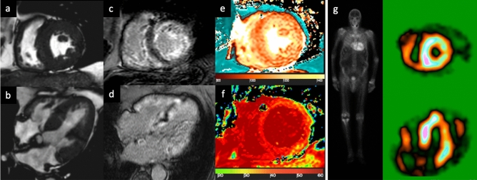
Fig. 2.
83-year-old male patient with known CAD and the presence of dyspnoea. Cine-SSFP sequences (a short axis view; b 4-chamber view), which show a thickening of both the left ventricular myocardium (18 mm in the septum) and the right ventricle, but also of the atrial walls with global and severe hypokinesia (left ventricular ejection fraction 26%). Inversion recovery turbo field echo sequences (c short axis view; d 4-chamber view) for late gadolinium enhancement (LGE) analysis; there are diffuse areas of circumferential subendocardial pattern enhancement even with transmural extension in the basal segment. There is also LGE within the right ventricle and both atrial walls. The quantitative evaluation of global left ventricular myocardium native T1 (e short axis view) and ECV (f short axis view) resulted in 1110 ms (v.n. 1000 ms) and 55% (v.n. 20–30%), respectively. Overall, the presence and the pattern of LGE with a transmural pattern in both ventricle and atrial walls were suspicious of transthyretin (ATTR) amyloidosis. The patient was then scanned with 99mTc-DPD (image g), where the abnormal and diffuse presence of the osteotropic indicator is observed in the left and right ventricle with a Perugini score = 3. The final diagnosis was ATTR amyloidosis

