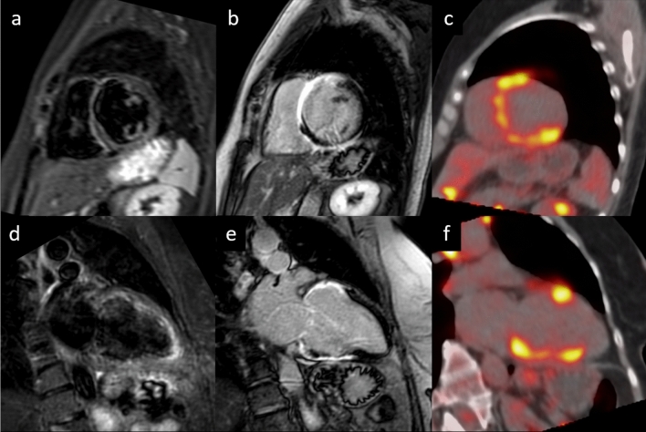
Fig. 3.
57-year-old female with frequent syncopal episodes and ventricular tachycardia, LV dilation and severe reduction in EF at TTE, with no obstruction of coronary arteries at coronary angiography. CMR revealed no edema on T2w-STIR images (a short axis view and d LV long axis view) and extensive areas of late gadolinium enhancement at IR-TFE images (b short axis view and e LV long axis view), with a non-ischemic pattern of distribution. The FDG-PET (c short axis view, f long axis view) confirmed the diagnosis of sarcoidosis with the identification of areas of FDG uptake (i.e., active inflammation) within the myocardium and in the mediastinal lymph nodes. LV left ventricle, EF ejection fraction; TTE transthoracic echocardium

