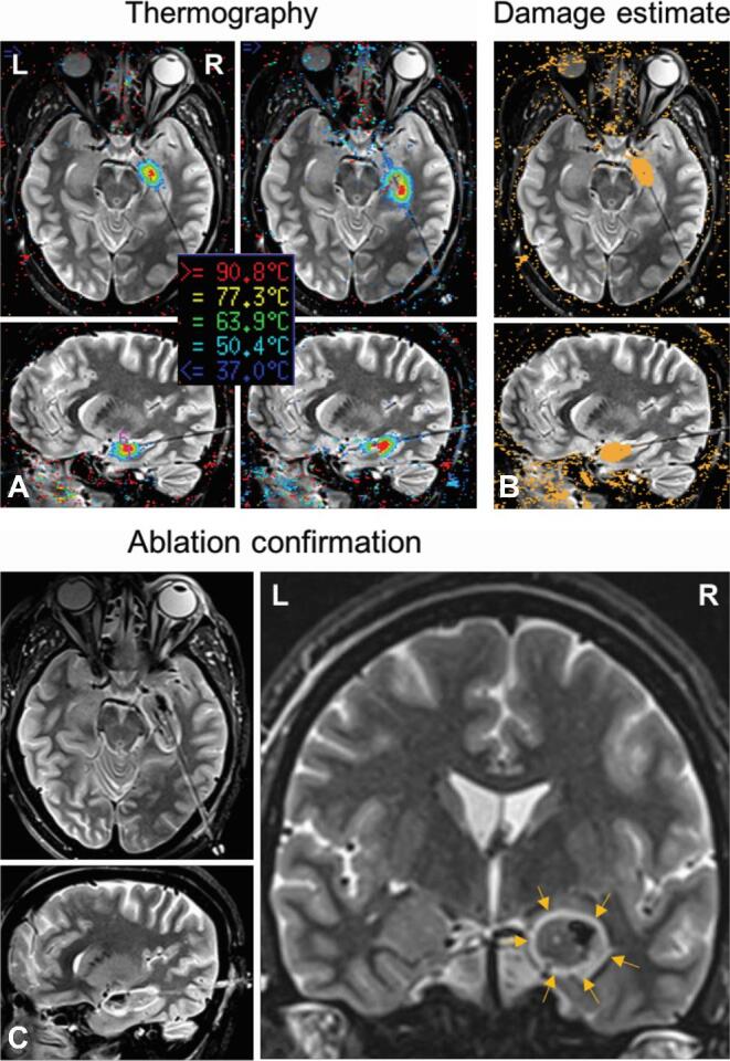
FIGURE 2.

Stereotactic laser amygdalohippocampotomy in patient 2. A, Thermography views demonstrate probe location in approximately axial and sagittal trajectory views of T2-weighted inversion-recovery-sequence MRI. Coregistered thermographic heat maps display sequential real-time ablation of right amygdala and anterior hippocampus along a length of the laser probe. B, Damage estimates demonstrate cumulative calculated irreversible damage zones (orange areas) for individual ablation locations in each trajectory plane. C, Ablation confirmation (T2-weighted inversion-recovery) shows the extent of final ablation in trajectory and standard coronal views. Arrows demarcate the rim of hyperintense edema surrounding the ablation zone.
