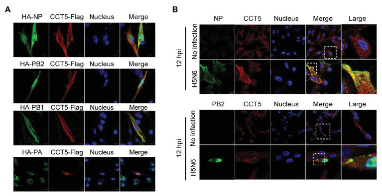
Figure 4.
Colocalization of nucleoprotein (NP), polymerase basic protein 2 (PB2), and polymerase basic protein 1 (PB1) with CCT5 protein. (A) CCT5-Flag and HA-NP, HA-PB2, HA-PB1, or HA-PA were cotransfected into DF-1 cells for 24 h. CCT5-Flag and HA-NP, HA-PB2, HA-PB1, or HA-PA were labeled with anti-Flag mouse and anti-HA rabbit antibodies and stained with antimouse 594 red fluorescent secondary antibody and 488 green fluorescent antirabbit antibodies, respectively. The nucleus was stained with 4’,6-diamidino-2-phenylindole (DAPI). The red, green, blue, and yellow fluorescence represents the CCT5-Flag, the HA-NP, HA-PB2, HA-PB1, or HA-PA, the localization of the nucleus, and the colocalization region, respectively. (B) DF-1 cells were infected with or without the H5N6-WT virus, and then, anti-CCT5 rabbit red fluorescent antibody and anti-NP/PB2 mouse green fluorescent antibody were used for immunostaining. The white boxed region was enlarged, as shown in the right. These experiments were examined with a confocal microscope (LSM 880; Zeiss) from three independent experiments.

