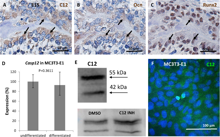
FIGURE 4.
Expression of caspase-12 in osteoblasts. Comparison of serial sections of the mouse mandibular bone at the prenatal/embryonic (E) day 15 showing co-localization of caspase-12 (A) with osteoblastic markers osteocalcin (B) and Runx2 (C) within the bone. Positive cells are brown, counterstained by hematoxylin (blue). Arrows point to cells in the same region. Scale bar: 50 μm. Expression of caspase-12 in undifferentiated and differentiated MC3T3-E1 cells (D) showing stable expression of Casp12 after 21 days of differentiation. Western blot analysis (E) and immunofluorescence (F) confirmed the presence of caspase-12 protein in MC3T3-E1. E: 55 kDa-full length caspase-12, 42 kDa-cleaved caspase-12, DMSO: caspase-12 forms after DMSO treatment, C12 INH: caspase-12 forms after specific inhibitor treatment. F: Caspase-12 in green, DAPI in blue. Scale bar: 100 μm.

