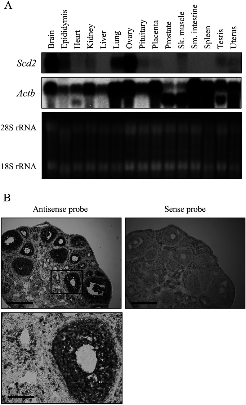
Fig. 1.
Mouse stearoyl-CoA desaturase 2 (Scd2) mRNA expression. (A) Northern blot analysis of Scd2 in various mouse tissues. Twenty micrograms of total RNAs from 15 tissues were electrophoresed on a denaturing agarose gel and stained with ethidium bromide (bottom). The gel was blotted to a nylon membrane and hybridized with a 32P-labeled Scd2 or Actb probe. The signals were detected by autoradiography (top and middle). The probe for Actb cross-hybridized to Actg1 and Actg2 due to high sequence homology, and the upper band is the signal of Actb and Actg1 and the lower band is that of Actg2. Intense Scd2 signal was observed in brain and ovary. (B) In situ hybridization analysis of Scd2 in the mouse ovary. Frozen sections (10 µm) from an ovary of a superovulated mouse were hybridized with a digoxigenin-labeled antisense or sense cRNA probe. A region marked in a box in the upper panel is shown at higher magnification at the bottom. The scale bar represents 400 µm in upper panels and 100 µm in a lower panel.

