
Figure 5.
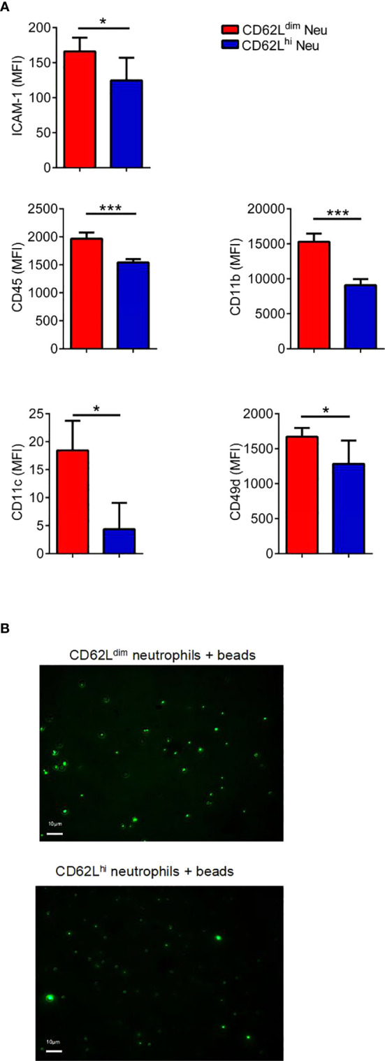
CD62Ldim neutrophils exhibit stronger adhesion. (A) MFI of adhesion molecules expressed on PB CD62Ldim and CD62Lhi neutrophils from 1-week tumor-bearing mice, as analyzed using flow cytometry. (B) Fluorescence intensity and images of residual fluorescent yellow-green latex beads adhered to CD62Ldim and CD62Lhi neutrophils. Data are presented as the mean ± SEM of one representative experiment. Similar results were obtained in three independent experiments. Unpaired Student’s t tests, ns, not significant. *p < 0.05 and ***p < 0.001.