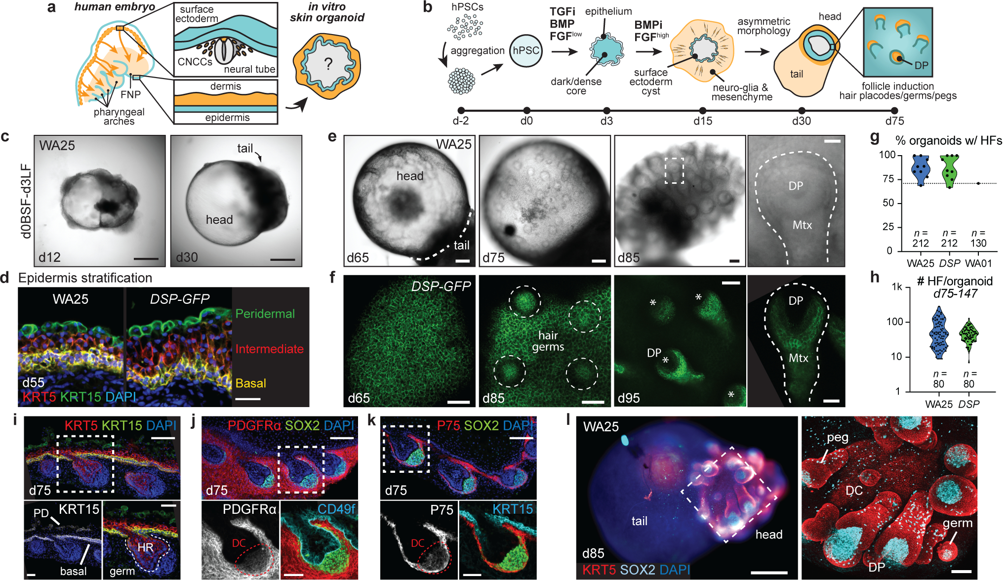
Figure 1 |. Surface ectoderm and CNCC co-induction leads to hair-bearing skin generation.

a, b, Overview of (a) study objectives and (b) skin organoid (SkO) protocol. c, Brightfield images of WA25 aggregates on days 12 and 30 in optimized culture. d, Immunostaining for KRT5+KRT15+ basal and KRT15+ peridermal layers at day-55. e, f, Representative HF-induction images (e) in brightfield of days 65–85 WA25 SkOs and (f) max-intensity confocal image (endogenous DSP-GFP) of days 65–95 DSP-GFP SkO. Dashed-box: magnified-HF; dashed-line: HF; dashed-circles: developing hair germs; asterisks: dermal papilla. g, h, Violin plots showing (g) frequencies of HF-formation in WA25 (average 87.4%, min=68.8%, max=100%, n=212 organoids), DSP-GFP (average 87.2%, min=66.7%, max=100%, n=212 organoids), and WA01 (71%, n=130 organoids) cultures, and (h) average number of HFs formed in WA25 (average 64 HFs/organoid, min=9, max=285, n=80 organoids) and DSP-GFP (average 48 HFs/organoid, min=7, max=128, n=80 organoids) cultures between days 75–147. i-k, Immunostained day-75 WA25 SkO with hair placodes. Antibodies highlight epidermal (KRT5+KRT15+CD49f+) and periderm (KRT15+) layers, dermis (PDGFRα+P75+), and DC cells (SOX2+PDGFRα+P75+). Dashed-boxes: magnified-regions. l, Wholemount of day-85 WA25 SkO with head-tail structures. KRT5 highlights epidermis and HF outer root sheath. SOX2 marks DC, DP, Merkel cells and melanocytes. Dashed-box: area shown to the right. Abbr: frontonasal prominence (FNP); cranial neural crest cells (CNCCs); dermal papilla (DP); matrix (Mtx); periderm (PD); hair root (HR); dermal condensate (DC). Scale: 500 μm (c), 250 μm (l; left), 100 μm (e; first three panels, f; third-panel, i-k; upper-panels), 50 μm (d, f; second-panel, i-k; lower-panels, l; right), 25 μm (e; last-panel, f; first/last-panels). See Statistics and Reproducibility for plot and experimental information.
