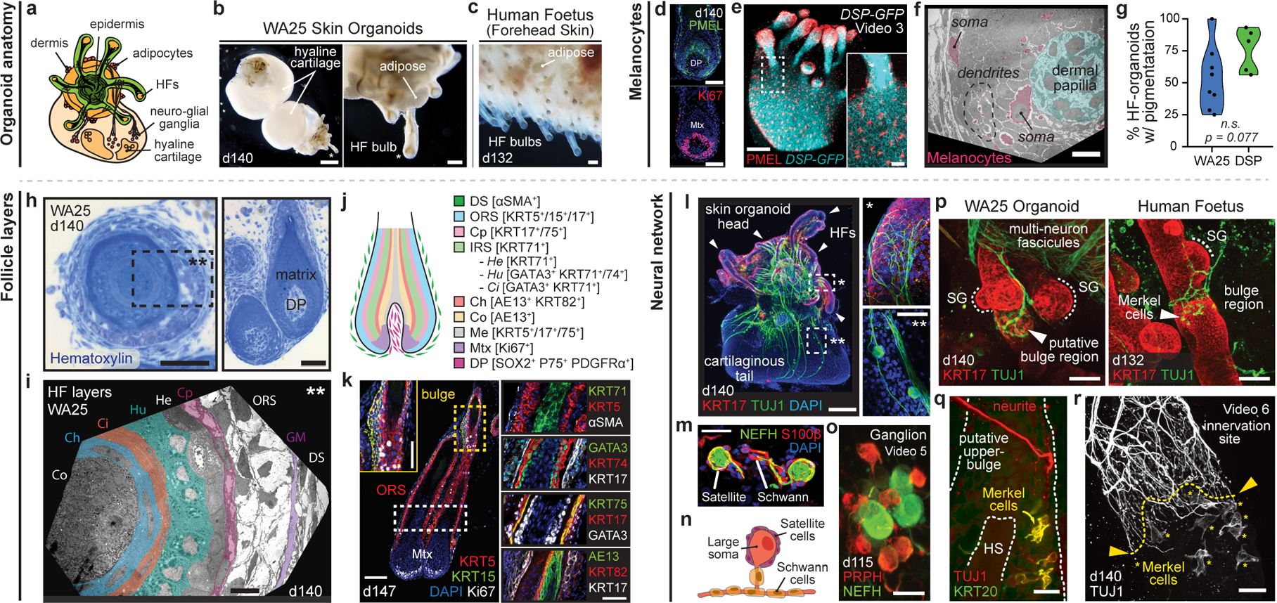
Figure 3 |. Skin organoid HF pigmentation, structure, and neural innervation.

a-c, Skin organoid (SkO) anatomy: (a) Schematic; typical SkO. (b, c), Darkfield-images; (b) day-140 WA25 SkOs; cartilage in organoid-tail and (c) 18-week human foetal skin. d-g, Melanocytes: (d) PMEL/Ki67 immunostaining; WA25 organoid-HFs and (e) PMEL+ melanocytes in DSP-GFP organoid-HFs and -epithelium. (f) Transmission electron microscopy (TEM); matrix-associated melanocytes (pink). (g) Violin plots compare HF-pigmentation frequency between WA25 (average=53.5%, min=25%, max=100%, n=137 organoids) and DSP-GFP (average=76.2%, min=56.3%, max=93.3%, n=91 organoids); Welch’s two-sided t-test, p=0.077. h-k, HF-layers: (h) Hematoxylin-staining; day-140 WA25 HFs. Dashed-box: region in (i) TEM; HF-layers. (j) Schematic; human HF-layers. (k) Immunostaining for markers in (j) on day-147 organoid-HFs; each layer from outermost ⍺SMA+ DS to innermost AE13+ Co. l-r, Neural network: (l) KRT17/TUJ1 wholemount; neurons in day-140 WA25 SkO. Magnified-area with asterisks: neurites targeting (*) HF-bulge and (**) neuron-soma. Arrowheads: HF-clusters. (m) Neurofilament-heavy chain (NEFH)-positive neurons with S100β+ satellite glial and Schwann cells. (n) Schematic; typical SkO-neurons. (o) Wholemount of day-115 WA25 SkO; ganglion with PRPH+NEFH− and PRPHLOWNEFH+ neurons. (p) Wholemount for TUJ1/KRT17; (left) day-140 WA25 SkO and (right) 18-week human foetus-forehead skin; neurites innervate HF-upper-bulge. HF-SGs noted. (q, r) TUJ1/KRT20 wholemount on day-140 WA25 SkOs; TUJ1+ neurites innervate HF-upper-bulge above TUJ1+KRT20+ (yellow) Merkel cells. (r) High-magnification of innervation site. Yellow-asterisks: Merkel cells. Abbr: hair shaft (HS); dermal sheath (DS); glassy membrane (GM); outer root sheath (ORS); companion (Cp); inner root sheath (IRS); Henle’s (He); Huxley’s (Hu); IRS-cuticle (Ci); cuticle (Ch); cortex (Co); medulla (Me); sebaceous gland (SG). Scale: 250 μm (b; upper, l; left), 100 μm (b; lower, c, e, k; left), 50 μm (d, h; right, k; insert and right-panels, p), 25 μm (e; insert, h; left, l; right-panels, m, o, q, r), 10 μm (f, i). See Statistics and Reproducibility for statistics and experimental information.
