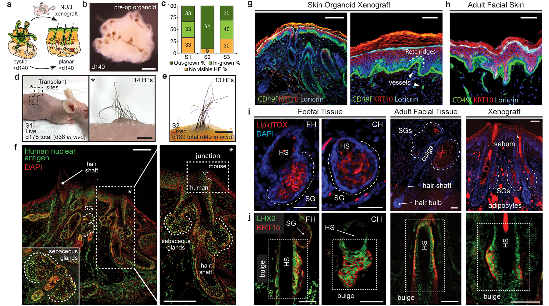
Figure 4 |. Skin organoids undergo cystic-to-planar transition when xenografted.

a, Schematic of skin organoid (SkO) xenografting strategy. b, Darkfield image of day-140 WA25 SkO prior to grafting (pre-operative; pre-op). c, Quantification of HF-growth percentage throughout xenograft experiments. Data compiled from 27 xenografts performed over 3 separate experiments/surgeries (S1, S2, S3). d, A xenografted NU/J mouse at 38-days post-operative (PO) following the first experiment (S1); total 178-days-old SkO. Pigmented hair shafts are visible at both graft sites, highlighted by boxes. One graft site (right) shows 14 HFs. e, S2 xenograft containing out-grown pigmented-hairs at 49-days PO; total 189-days-old SkO. f, Immunostaining for Human nuclear antigen and DAPI in out-grown xenograft. Note mouse and human epidermis junction (right). Organoid-HFs have SGs. g, h, Comparison of epidermis layers between (g) SkO-xenograft and (h) adult facial skin by immunostaining for CD49f (basal), KRT10 (intermediate), and Loricrin (granular). Unspecific staining above Loricrin+ granular layer is cornified layer. Note Rete ridges in xenograft and adult skin. Arrowheads; vessels. i, j Comparison of (i) LipidTOX+ SGs and (j) LHX2+KRT15+ putative bulge stem cells at HF-bulge region in (left) foetal forehead and cheek, (middle) adult facial, and (right) xenograft. (i) LipidTOX also labeled adipocytes. Dashed-lines; SGs. (j) Dashed-boxes: HF-bulge. Abbr: operative (OP); forehead (FH); cheek (CH). Scale: 1 mm (d; right, e), 200 μm (b, f, g; left), 100 μm (i & j; Adult & Xenograft), 50 μm (g; right, h, i & j; Foetal). See Statistics and Reproducibility for experimental information.
