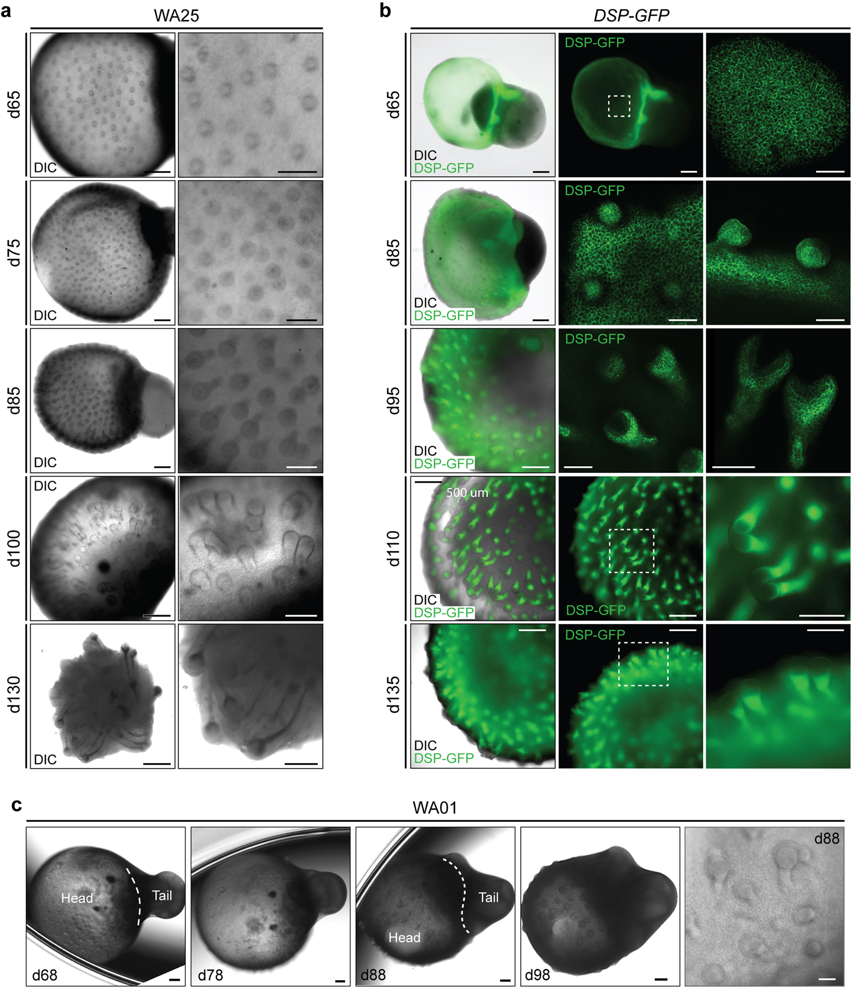
Extended Data Figure 3 |. Comparison of initial hair follicle induction in WA25 and DSP-GFP live-cell aggregates.

a, Representative DIC images of days 65–130 WA25 skin organoids with developing HFs. b, DIC and endogenous GFP fluorescence images of days 65–135 DSP-GFP skin organoids with developing HFs. c, DIC images of days 68–98 WA01 skin organoids with developing HFs. Magnified view of day-88 WA01 skin organoid HFs is shown in the last panel. Note that the head (skin cyst)-and-tail (non-skin mesenchymal) portions within skin organoids are distinguishable. Also, note that the hair bulbs are facing outward of the organoid, contacting medium, while the hair shafts grow inward toward the center of the skin organoid. Dashed boxes indicate magnified regions. Scale bars: 500 μm (a; left panels, b; left/middle panels), 250 μm (a & b; right panels, c; all panels except last panel), 100 μm (b; d65 & 85 right panels), 50 μm (c; last panel). WA25 and DSP-GFP skin organoid images represent morphologies observed throughout 9 independent cultures, and WA01 skin organoid images represent one experiment performed by the Stanford group. Corresponds with data in Fig. 1e and f.
