. 2020 Feb 24;11(3):392–410. doi: 10.1039/c9md00594c

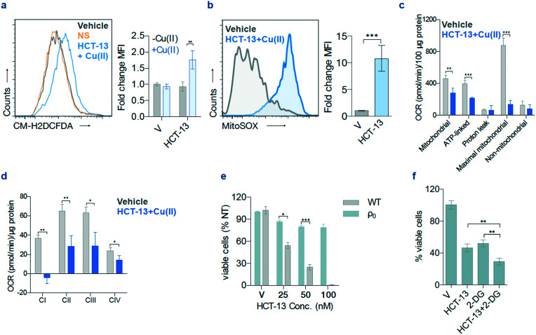
Fig. 5. HCT-13 + Cu(ii) induces oxidative stress and has selective mitochondrial toxicity. (a) Reactive oxygen species (ROS) measurements using CM-H2DCFDA after HCT-13 (25 nM) ± Cu(ii) (20 μM) treatment for 24 h; mean fluorescence intensity (MFI); mean ± SD, n = 2, Student t-test, ** P < 0.01. (b) Mitochondrial ROS detection using MitoSOX in MIAPACA2 PDAC cells treated with HCT-13 (25 nM) + Cu(ii) (20 μM) for 24 h. mean ± SD, n = 2, Student t-test, *** P < 0.001. (c) Oxygen consumption rate (OCR) of MIAPACA2 PDAC cells treated with HCT-13 (25 nM) + Cu(ii) (20 μM) for 24 h; ** P < 0.01; *** P < 0.001. (d) OCR of isolated mitochondria measured with or without HCT-13 (25 nM) + Cu(ii) (20 μM); * P < 0.05; ** P < 0.01. (e) Viability of 143 BTK parental (wild type, WT) and ρ0 cells after 48 h of the indicated HCT-13 concentration + Cu(ii) (20 μM) treatment, assessed with trypan blue staining; mean ± SD, n = 2, Student t-test, * P < 0.05; *** P < 0.001; V: vehicle. (f) Cell viability by trypan blue staining in MIAPACA2 PDAC cells to interrogate the interaction of HCT-13 (25 nM) + Cu(ii) (20 μM) with 2-DG (2 mM) for 48 h; mean ± SD, n = 2, Student t-test, ** P < 0.01; V: vehicle.

Fig. 5