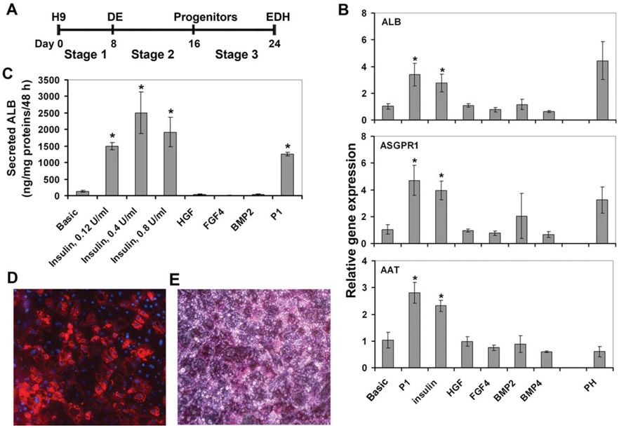
Figure 1.
Insulin enhanced hepatocyte differentiation. (A): Schematic representation of the differentiation protocol. (B): Quantitative RT-PCR analysis of hepatic gene expression in EDHs cultured with indicated growth factors at stage 2. *, p<.005 versus Basic. (C): Secreted ALB levels in the culture media were assessed by ELISA. *, p<.005 versus Basic. (D): Immunofluorescent staining of EDHs derived using the P2 protocol for ALB. Magnification: 200. (E): Periodic acid Schiff stain of EDHs derived using the P2 protocol. Magnification: 200. Abbreviations: DE, definitive endoderm; EDH, embryonic stem cell-derived hepatocyte.

