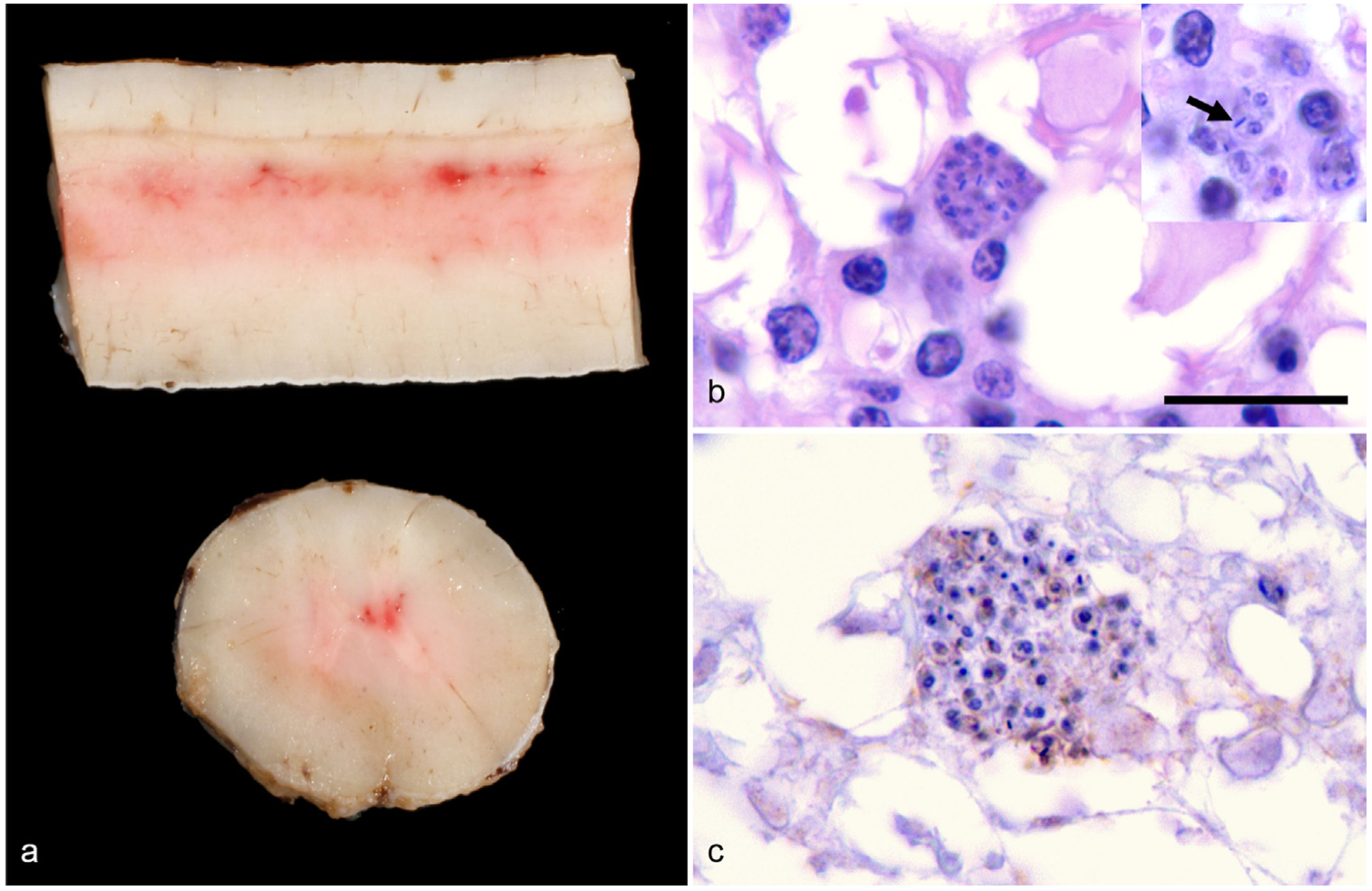
Fig. 1.

Gross and histologic lesions within the spinal cord of a 10-year-old Quarter Horse gelding from Texas. (a) Longitudinal and transverse sections of the thoracic spinal cord reveal multifocal areas of congestion and mild hemorrhage within the dorsal and ventral horn gray matter. (b) Histologically, clusters of 2–3 μm in diameter, protozoan amastigotes surrounded by a few plasma cells are in the hyperemic areas of spinal cord identified in panel a. In the insert, amastigotes have a thin outer membrane, contain a 1 μm basophilic nucleus and have a bar-shaped kinetoplast (arrow) that is often oriented parallel to the nucleus; HE, 1000×, Bar = 10 μm. (c) Large clusters of amastigotes are negative with Sarcocystis spp.—specific immunohistochemical staining, which would stain organisms bright red if positive; IgG antibody conjugated with horseradish peroxidase, 1000×.
