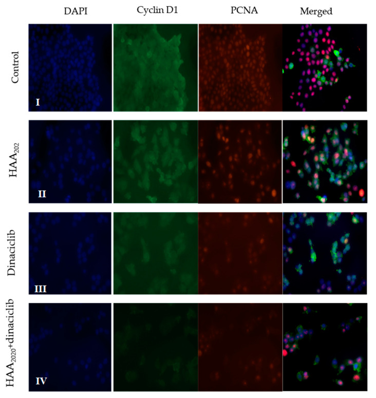
Figure 9.
The co-localization of the immunofluorescent bodies using cyclin D1 (green) combined with PCNA (red), counterstained with DAPI (4‘,6-diamidino-2-phenylindole) in MCF7 cells (scale bar = 15 μm; 40× objective). I: untreated control, II: cells treated with HAA2020, III: cells treated with dinaciclib, IV: cells treated with both HAA2020, dinaciclib.

