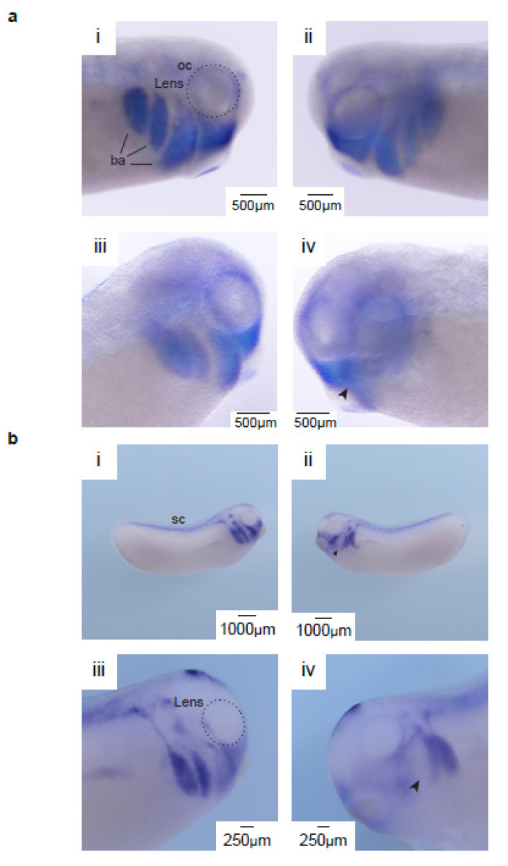
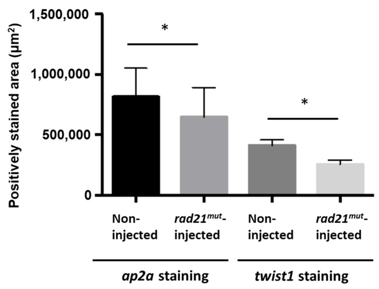
Figure 3.
rad21mut disrupts the migration of neural crest cells (NCCs) in stage-25 Xenopus laevis. (a) The in situ hybridization pattern of twist1 was altered in the rad21mut-injected side. Panel i and ii show twist1 expression on both sides of the same non-injected embryo. The patterns around the optic cup (oc) and the neural crest migration streams into branchial arches (ba) are intact (indicated by a dash circle and dark solid lines, respectively). Panel iii and iv show the non-injected side and the injected side of the same rad21mut-injected embryo. The optic cup and branchial arch in panel iii are similar to those in panel i and ii, whereas these patterns are disrupted in panel iv. A total of 20 rad21mut-injected embryos were used, and 18 of them showed disrupted patterns in the rad21mut-injected side (18/20). (b) The in situ hybridization pattern of ap2a is altered in rad21mut-injected X. laevis embryos. Panel i shows ap2a is highly expressed in the head region and the spinal cord (sc) in the non-injected side. Panel ii shows the ap2a expression pattern is disrupted in the rad21mut-injected side. A total of 11 rad21mut-injected embryos were used, and 9 showed disrupted staining patterns in the rad21mut-injected side (9/11, arrowhead). Panel iii and iv show higher magnifications of the non-injected side and rad21mut-injected side of another embryo. Similar to panel ii, disrupted neural crest migration streams are shown in panel iv. A total of 11 rad21mut-injected embryos were used, and 9 showed disrupted staining patterns in the rad21mut-injected side (9/11, arrowhead). (c) Quantification of the mandibular areas showing positive staining of ap2a and twist1. Means and standard deviations for three injected sides were analyzed in each group. Statistics was done by using paired t test; * indicates p < 0.05.


