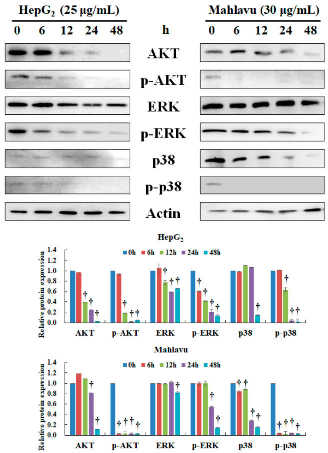
Figure 3.
Effect of CAt extract treatment on AKT, ERK, and p38 protein expression in HepG2 and Mahlavu cells. HepG2 and Mahlavu cells were incubated with CAt extract (25 or 30 μg/mL) for the indicated time points (0, 6, 12, 24, and 48 h) then subjected to Western blotting analysis. The actin level was used as loading control. †: Significant difference between control and treatment, p < 0.05.

