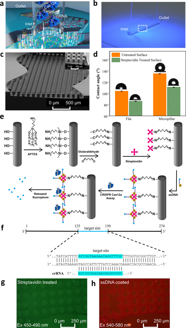
Figure 1.
(a) Schematic of the clustered regularly interspaced short palindromic repeats (CRISPR)-based IMPACT chip DNA detection. (b) Photograph of the IMPACT chip. The dashed white box indicates regions patterned with micropillars. (c) SEM image of the micropillars. Inset: high magnification image. (d) Static water contact angle measurement on both flat and micropillar surfaces before (orange) and after (green) surface treatment. (e) Schematic of the surface treatment protocol, ssDNA probe binding, and CRISPR detection. (f) ASFV target DNA sequence and the corresponding crRNA sequence. (g) Fluorescent image of the channel which received chemical treatment and streptavidin binding (green fluorescence). (h) Fluorescent ssDNA-covered IMPACT chip, showing red fluorescence.

