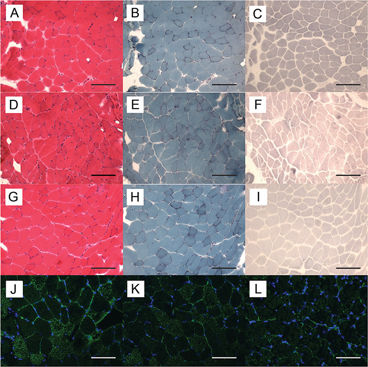
Figure 3.

Histological and immunohistochemical findings in deltoid muscle of female mice. Representative serial cryosections of deltoid muscle of a WT (A–C), Het (D–F) and Homo female mice (G–I) at age 10 months. (A, D and G) hematoxylin-eosin stain; (B, E and H) modified Gomori-Trichrome stain; (C, F and I) menadione-NBT stain. Bar = 75 μm. There were no differences in staining intensities or structural abnormalities. Frozen sections were stained with anti-Fhl1 antibody (green) and DAPI (blue). Representative immunohistochemistry of deltoid muscle of a WT (J), a Het and (K) and a Homo female mouse (L) at age 20 months. Bar = 75 μm. Homo female mouse showed decreased Fhl1 signal intensity compared to WT and Het female mice.
