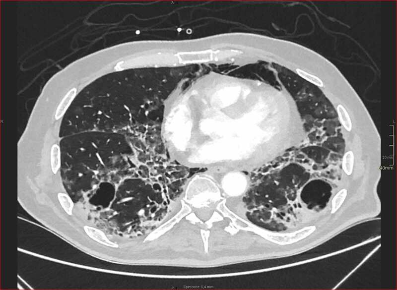
Figure 1.

High-resolution lung base image from contrast-enhancement arterial scan for pulmonary embolism detection in patient with long-standing COVID-19 pneumonia and pneumomediastinum. Ground-glass opacities are detected in subpleural areas mixed with focal consolidations. Moreover, computed tomography shows initial fibrotic changes with architectural distortion and bronchiolectasis. Multiple thin-walled cysts are also recognized, in keeping with smoking-related changes (courtesy by Gabriele D’Andrea, Radiology Unit, San Gerardo Hospital, ASST Monza, Monza, Italy).
