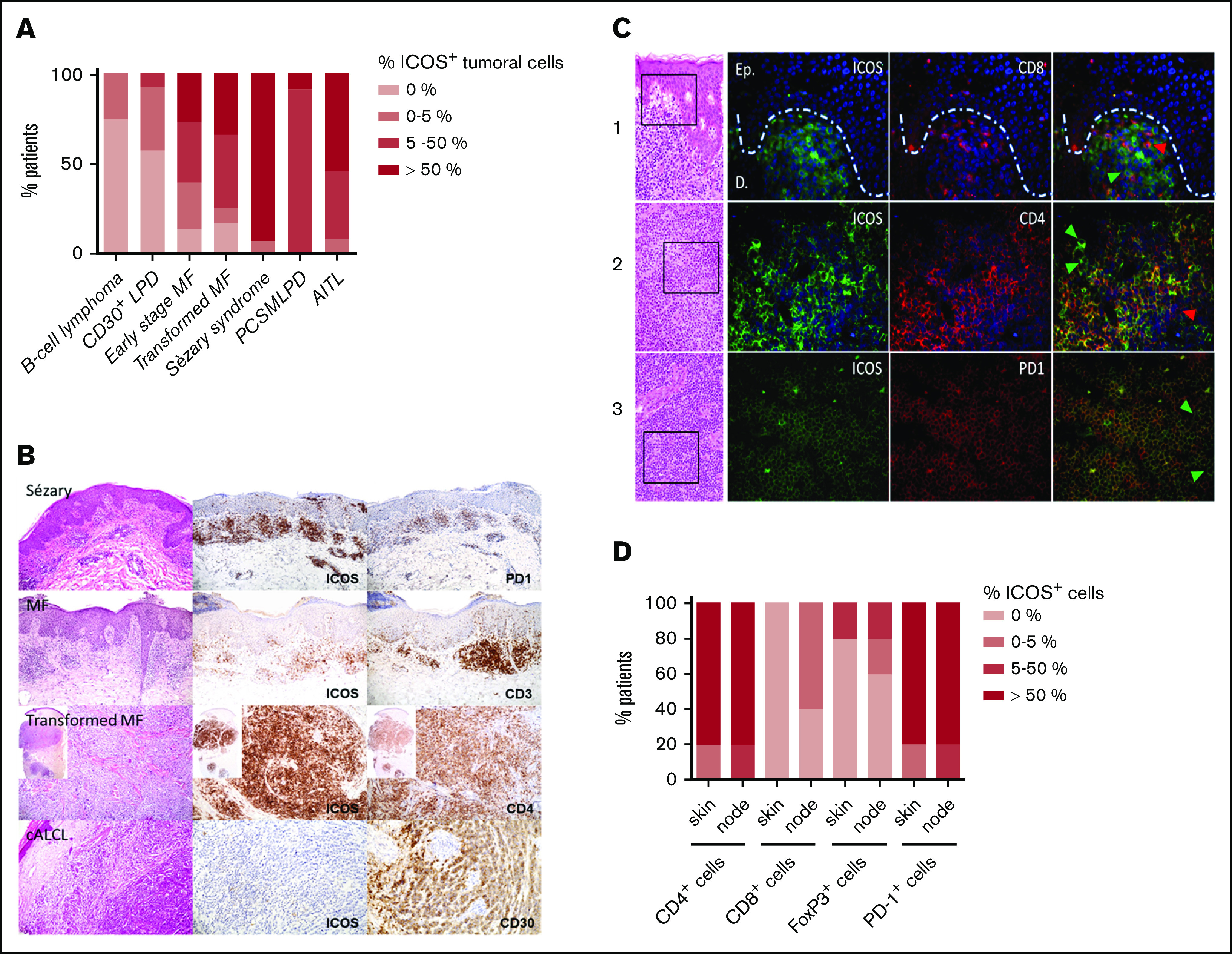
Figure 1.

ICOS expression by malignant T cells in CTCL skin biopsy specimens at different disease stages. (A) Percentage of patients with ICOS+ tumoral cells, determined by immunohistochemical analysis of ICOS expression in skin samples of 23 patients with early-stage MF, 12 with transformed MF, 17 with SS, 12 with B-cell lymphoma, 14 with CD30+ LPD (cutaneous anaplastic large cell lymphoma and lymphomatoid papulosis), 12 with PCSMTLPD, and 13 with AITL. Cutaneous B-cell lymphoma skin samples were used as negative controls, whereas PCSMTLPD and AITL were positive controls. (B) Immunohistochemical analysis of the expression of ICOS in epidermal or dermal CD3+CD4+ infiltrating lymphocytes. (C) Double-staining experiments using immunofluorescence with anti-mouse Texas red anti-rabbit Alexa Fluor 488 as secondary antibodies and 4′,6-diamidino-2-phenylindole for nuclear staining in skin (line 1) and lymph node samples (lines 2 and 3) from 3 different patients with SS. Arrowheads identify cells with a single-marker expression; green arrowheads show ICOS+ cells, and red arrowheads show CD8+ cells (line 2) and CD4+ cells (line 3). (D) Double-staining immunohistochemical analysis of skin and node samples of 5 patients with SS. The percentage of patients with ICOS+/CD4+, ICOS+/CD8+, ICOS+/FoxP3+, and ICOS+/PD-1+ cells in the skin and node tumoral infiltrate is represented. cALCL, cutaneous anaplastic large cell lymphoma.
