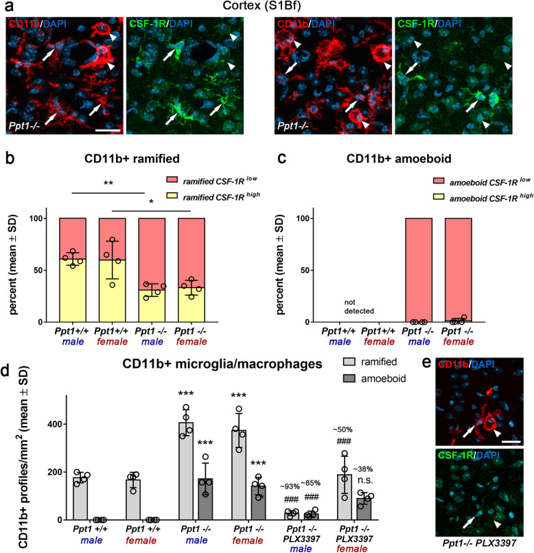
Fig. 3.
High levels of CSF-1R expression correlate with efficient depletion of CD11b+ cells in the cortex of CLN1 mice. a Representative fluorescent microscopic images of immunohistochemical labeling of CSF-1R on CD11b+ microglia/macrophages in the S1Bf cortex of untreated 6-month-old Ppt1−/− mice. Arrows: ramified (and hyper-ramified) cells; arrowheads: amoeboid cells. Scale bar: 20 μm. b, c Quantification of CSF-1R immunoreactivity on ramified and amoeboid CD11b+ microglia/macrophages in Ppt1+/+ and Ppt1+/+ mice showed that high CSF-1R expression was restricted to ramified CD11b+ cells. In addition, the percent of ramified cells highly expressing the CSF-1R was significantly reduced in untreated Ppt1−/− compared with Ppt1+/+ mice. Two-tailed unpaired Student’s t test. *P < 0.05; **P < 0.01. d Quantification of ramified and amoeboid CD11b+ cells in Ppt1+/+, Ppt1−/−, and PLX3397-treated Ppt1−/− mice showed that in mutant mice ramified cells were depleted more efficiently compared with amoeboid cells. n = 4 male and 4 female mice per group. One-way ANOVA and Tukey’s post hoc tests. ***, ###P < 0.001. # significant difference to untreated Ppt1−/− mice of the same sex. e Representative fluorescent microscopic images of immunohistochemical labeling of CSF-1R on CD11b+ microglia/macrophages in the S1Bf cortex of a 6-month-old PLX3397-treated Ppt1−/− mouse. Arrow: ramified cell; arrowhead: amoeboid cell. Scale bar: 20 μm

