Fig. 5.
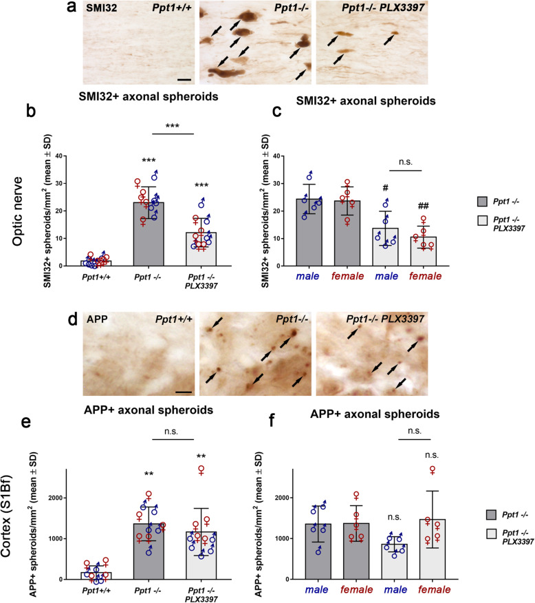
Depletion of microglia/macrophages attenuates axon damage in the CNS of CLN1 mice. a Immunohistochemical detection of SMI32+ axonal spheroids (arrows) in longitudinal optic nerve sections of 6-month-old Ppt1+/+, Ppt1−/− mice and PLX3397-treated Ppt1−/− mice. Scale bar: 20 μm. b Quantification of SMI32+ axonal spheroids optic nerves showed a significant reduction of ongoing axonal damage after microglia depletion. c Quantification of SMI32+ axonal spheroids of Ppt1−/− and PLX3397-treated Ppt1−/− mice separated by their sex did not show any differences regarding axonal spheroids. d Immunohistochemical detection of APP+ axonal spheroids (arrows) in the S1Bf cortex region of 6-month-old Ppt1+/+, Ppt1−/−, and PLX3397-treated Ppt1−/− mice. Scale bar: 20 μm. e Quantification of APP+ axonal spheroids in the S1Bf cortex region revealed that microglia/macrophage depletion did not significantly attenuate ongoing axonal damage within this CNS compartment. f Quantification of APP+ axonal spheroids in the cortex separated by sex showed a non-significant tendency to reduction of spheroid numbers in male but not female PLX3397-treated Ppt1−/− mice. n = 5 male and 5 female mice per group. One-way ANOVA and Tukey’s post hoc tests. #P < 0.05; **, ##P < 0.01; ***P < 0.001. # significant difference to untreated Ppt1−/− mice of the same sex

