Figure 2.
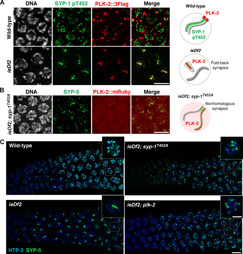
Constrained PLK-2 activity at the pairing centers is essential for proper synapsis. (A) Immunofluorescence images of early pachytene nuclei from wild-type and ieDf2 mutants stained for DNA (white), SYP-1 pT452 (green), and PLK-2::3Flag (red). Scale bar, 5 µm. (B) Immunofluorescence images of mid-pachytene nuclei from ieDf2; syp-1T452A mutants stained for DNA (white), SYP-1 (green), and PLK-2::mRuby (red). Scale bar, 5 µm. Diagrams illustrating the results are shown on the right. (C) Composite projection images of gonad sections from indicated genotypes, spanning from meiotic entry to mid-pachytene, showing HTP-3 (blue) and SYP-5 (green). Scale bar, 5 µm. Insets show zoomed-in view of a representative nucleus from the boxed regions. Scale bar, 2 µm.

