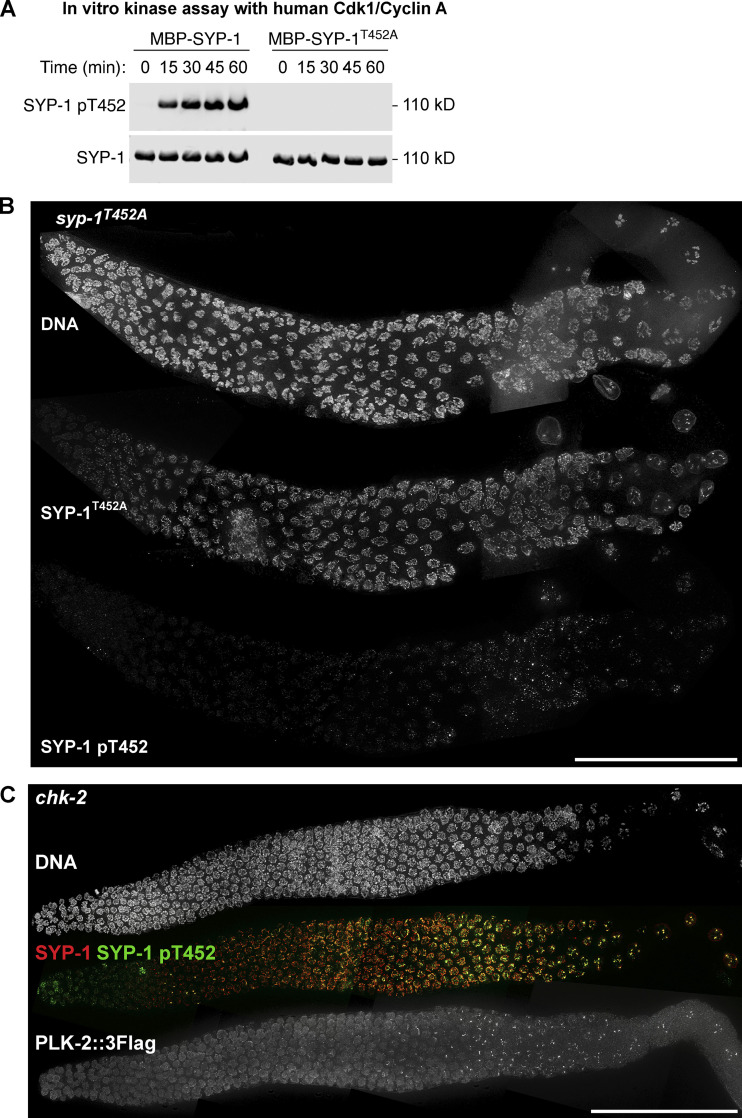
Figure S1.
Phospho-specificity of the SYP-1 pT452 antibody used in this study, and the analysis of SYP-1 T452 phosphorylation and PLK-2 localization in chk-2 mutants. (A) In vitro kinase assays using human CDK-1/cyclin A to phosphorylate MBP–SYP-1 with or without the T452A mutation. Western blot using antibodies against SYP-1 and phosphorylated SYP-1 at T452 is shown. (B) Composite immunofluorescence images of a whole gonad dissected from syp-1T452A mutants showing DNA, SYP-1, and SYP-1 pT452 staining. Scale bar, 50 µm. (C) Composite immunofluorescence images of a whole CHK-2–depleted gonad showing DNA (white), SYP-1 (red), SYP-1 pT452 (green), and PLK-2::3Flag staining. Scale bar, 50 µm.

