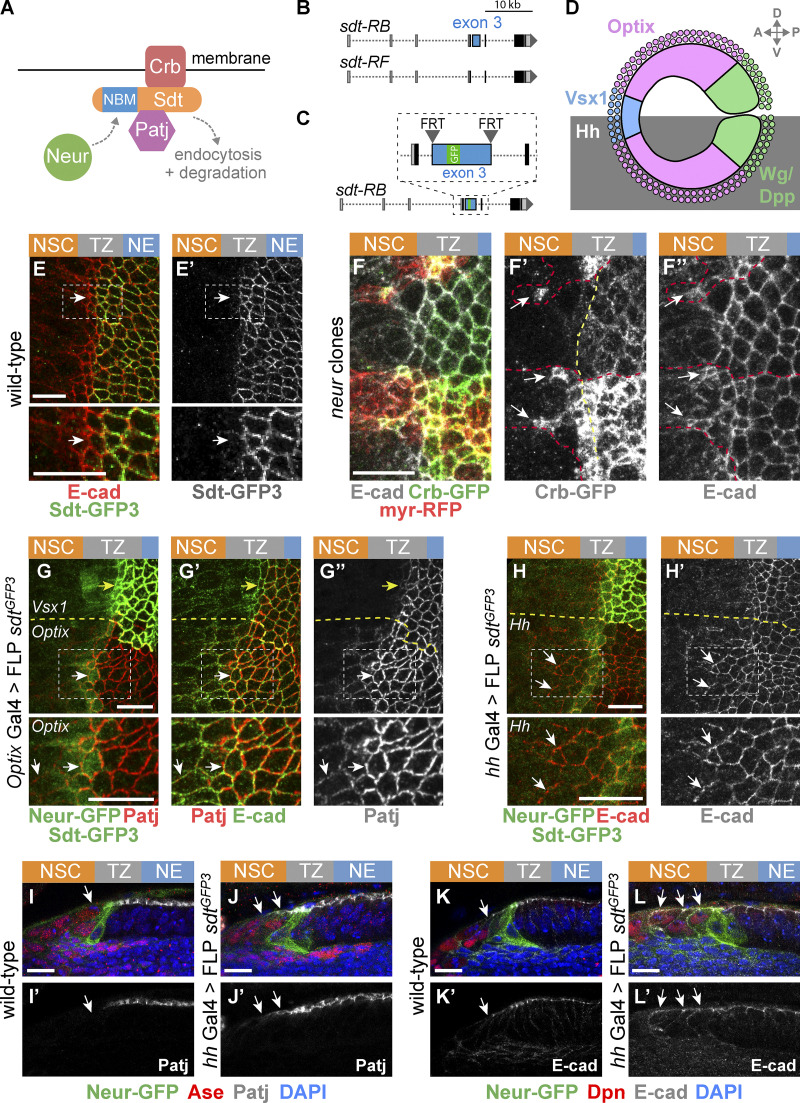
Figure 4.
Neur acts via Sdt to down-regulate Crb in epi-NSCs. (A) Working model of Crb regulation by Neur. Neur interacts directly with Sdt isoforms that include the NBM and targets the Crb complex for endocytosis and degradation. (B) Simplified view of the alternatively spliced sdt transcripts. A subset of the sdt mRNAs, such as RB but not RF, includes exon 3, which encodes the NBM of Sdt. (C) Molecular structure of the sdtGFP3 allele with GFP inserted into exon 3, itself flanked by FRT sites. (D) Diagram of the NE subdivided into spatial domains expressing different transcription factors including Optix and Vsx1, or signaling molecules including Dpp, Wg, and Hh, hence providing a toolbox of spatially restricted Gal4s. (E and E′) Sdt isoforms containing the sequence encoded by exon3 (Sdt-GFP3, green) were expressed in the NE and down-regulated in epi-NSCs (E-cad, red). (F–F″) Crb (Crb-GFP, green) was detected colocalizing with E-cad (white) in neur mutant cells past the TZ medial edge (arrows in F′ and F″). Mutant cells were marked by a membrane RFP (myrRFP, red) and had two copies of Crb-GFP, accounting for higher Crb-GFP levels. (G–G″) Expression of only Neur-resistant isoforms of Sdt, via the excision of exon 3 in the sdtGFP3 allele in the Optix domain (note the domain-specific loss of Std-GFP3, green in G), led to the detection of Patj (red in G) colocalizing with E-cad (green in G′) in mutant epi-NSCs (arrow; Neur-GFP, green in G) as well as in NSCs (arrowhead). In contrast, Patj was down-regulated in control epi-NSCs of the Vsx1 domain (yellow arrows). (H and H′) Excision of exon 3 of sdt in the Hh domain had a similar effect on E-Cad (red; see loss of Std-GFP3, green in G, in this domain; Neur-GFP, also green). (I–L′) Cross-section views showing that Patj (white in I–J′) was down-regulated in epi-NSCs (Neur-GFP, green) in the presence of Neur-sensitive Sdt in wild-type brains (I and I′) but not when only Neur-resistant Sdt was expressed upon excision of exon 3 in hh>FLP sdtGFP3 (J and J′). Note that Patj was observed at the apical cortex of NSCs (Ase, red) upon excision of exon 3 (J and J′). Likewise, E-cad (white in K–L′) was detected further within the NSC domain (Dpn, red) when Neur-resistant isoforms of Sdt were expressed (K–L′). DAPI (blue) marked all nuclei. Scale bars = 10 µm.

