Figure S3.
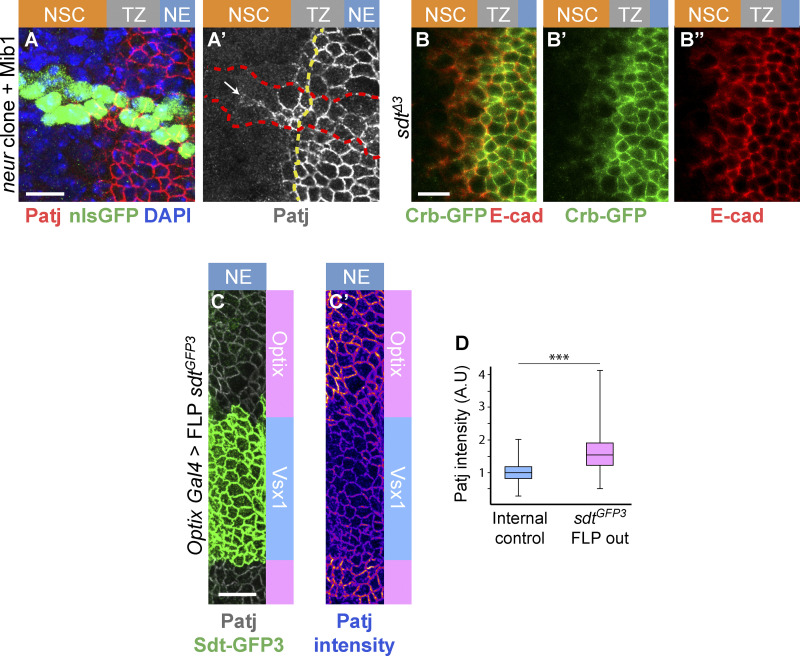
Analysis of the down-regulation of Crb and Patj. (A and A′) Patj (red) was observed to persist at the apical cortex of neurIF65 mutant cells overexpressing Mib1 (using tub-Gal4 in MARCM clones marked by nuclear GFP, green, outlined in red in A′; DAPI, blue) past the TZ medial edge. The boundary between epi-NSCs and other TZ cells is shown as a yellow dotted line. (B–B″) Crb-GFP (green; E-cad, red) was observed to persist at the apical cortex of the most medial TZ cells (epi-NSCs) in the OL of larvae carrying a deletion of exon 3 of sdt (sdtΔ3). (C and D) Loss of the NBM-containing isoforms of Sdt in the Optix domains (marked by the loss of Sdt-GFP3, green, upon FLP-mediated excision of exon 3) resulted in slightly higher accumulation of Patj (white, B; intensity code, B′), as quantified in D (n = 4). Scale bars = 10 µm. ***, P < 10−3.

