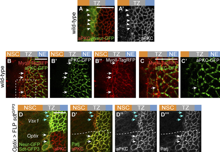
Figure S5.
Regulation of apical constriction by Neur and RhoGEF3. (A and A′) Low levels of aPKC (red) were detected in epi-NSCs (Neur-GFP, green) in wild-type brains. (B–C′) Snapshot of a living brain showing that aPKC-GFP (green) localized uniformly at apical junctions of NE and TZ cells but appeared to localize in patches at the apical cortex of epi-NSCs. Note that MyoII-TagRFP (red) was observed to be enriched along junctions where aPKC were decreased (white arrows). The inset boxed in B is shown in C and CRNAis. (D–D″′) While low aPKC levels were observed at the apical cortex of control epi-NSCs in the Vsx1 domain (blue arrows), expression of the Neur-resistant isoforms of Sdt (upon excision of exon 3 of sdt in the Optix domain, hence the loss of Sdt-GFP3, green) led to the persistent accumulation of aPKC (red; white arrows in L–L″) and Patj (green; LRNAis and L″) in epi-NSCs (Neur-GFP, green in L). In the Vsx1 domain, Sdt-GFP3 is down-regulated in epi-NSCs (blue arrows). Scale bars = 10 µm.

