Figure 2.
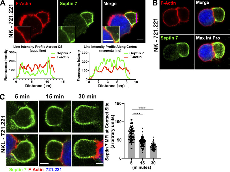
Septin 7 accumulates around the cell cortex and away from the CS in NK cells. (A) Primary NK cells were allowed to form conjugates for 15 min with CMAC-labeled 721.221 cells (blue) and stained for F-actin (red) or septin 7 (green) and imaged using confocal microscopy. Scale bar, 5 µm; inset scale bar, 2.5 µm. Line intensities profiles were created from the aqua line at the CS and magenta line along the NK cell cortex in images from A using Fiji. (B) NK cell–721.221 conjugates were formed for 15 min, and a z-stack image was obtained for septin 7 (green) and F-actin (red). The individual image shown is slice 6 of 19. (C) NK cell–721.221 conjugates were formed at the indicated time points and imaged for septin 7 (green) and F-actin (red). The MFI for septin 7 was measured in 60 conjugates (30 conjugates per time point in two separate experiments) across the CS using ImageJ. The data are graphed and shown to the right of the representative time point images. ****, P < 0.0001. P value indicated is for one-way ANOVA with Dunnett’s multiple comparison.

