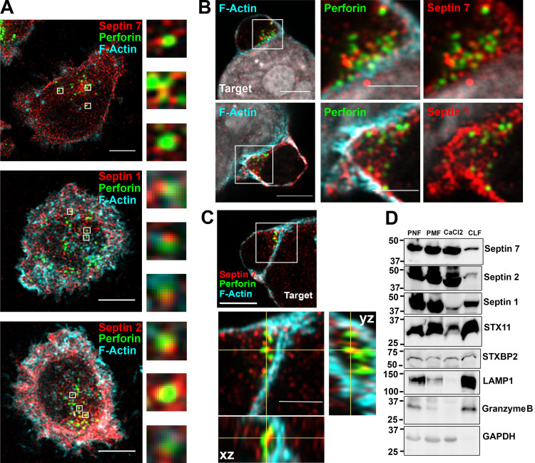
Figure 5.
Septins localize with lytic granules at the CS. Primary NK cells or their conjugates with 721.221 cells were stained for F-actin (cyan), septin 7, 1, or 2 (red), and perforin (green) to mark lytic granules. Cells were imaged using a confocal microscope in the Airyscan setting. (A) Septins were found closely apposed with perforin-containing lytic granules. Representative z-stack images are shown for septin 7 (top; slice 5 of 11), septin 1 (middle; slice 3 of 12), and septin 2 (slice 6 of 12). Scale bar, 5 µm. Magnified images are ∼1 µm. (B) Accumulation of septin 7 and septin 1 near lytic granules at the CS. Representative z-stack images are shown for septin 7 (top; slice 19 of 39) and septin 1 (bottom; slice 12 of 20). Scale bar, 5 µm; inset scale bar, 2.5 µm. (C) Co-localization of septin 7 with lytic granules at the CS shown in the Airyscan z-stack orthogonal views (slice 14 of 44). Scale bar, 5 µm; inset scale bar, 2.5 µm. (D) Postnuclear (PNL), postmitochondrial (PML), CaCl2, and CLF were isolated from unstimulated NKL cells as described in the Materials and methods section and immunoblotted as indicated. Immunoblot is representative of three independent experiments.

