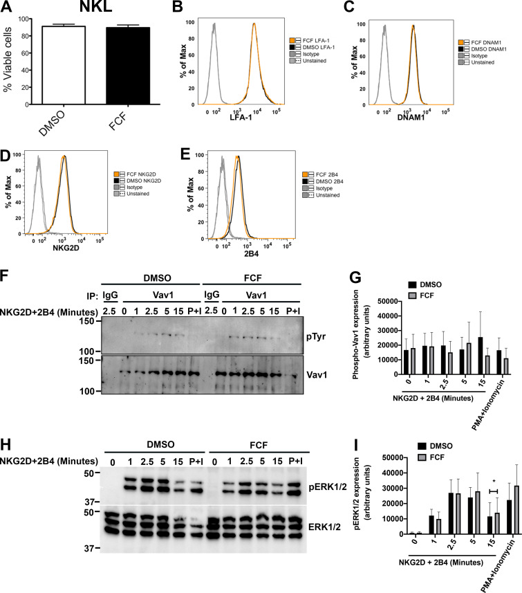
Figure S3.
Stabilization of septin filaments does not affect cell viability, surface expression of activating receptors, and signaling. NKL cells were treated with DMSO or 75 µM FCF for 3 h. (A) Cell viability was assessed. Results shown are the average of three independent experiments. Error bars indicate SEM. (B–E) Surface expression of activating receptors was measured using FC. Flow charts shown are representative of two independent experiments, each performed in triplicate. (F–I) NKL Cells were stimulated over the indicated time course by ligation of NKG2D/2B4 receptors or with PMA (2 µM) and ionomycin (4 µM; P+I) for 15 min. (F) Cell lysates were immunoprecipitated (IP) with anti-Vav1 or rabbit IgG, and activated Vav1 was detected by immunoblotting with anti-phosphotyrosine (pTyr) antibody. (G) Phospho-Vav1 bands were quantified using Bio-Rad Image Lab software and normalized to Vav1 expression. (H) Activated cells were immunoblotted as indicated. (I) Phospho-ERK1/2 (pERK1/2) bands were quantified and normalized to ERK1/2 expression. Blots shown are representative of three independent experiments. Error bars indicate SEM. *, P < 0.05. P values indicated are for paired, two-tailed Student’s t test.

