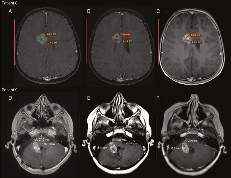
Fig. 2.
(A–C) Patient 8: post-contrast T1 weighted MR images show irregular intra- and periventricular enhancing tumor in the right frontal lobe. (A) Pre-infusion tumor size and characteristics. (B) After the fifth infusion the tumor shows irregular ring enhancement with central necrosis. (C) Ten days following infusion the tumor continues to minimally decrease in size with increased enhancement. (D–F). Patient 9: post-contrast T1 weighted images through posterior fossa show enhancing medulloblastoma centered along the right foramen of Luschka to the pontomedullary cistern with invasion of adjacent pons and right middle cerebellar peduncle. (D) Post-contrast T1 image before NK cell infusion. (E) Post-contrast image after 2 NK cell infusions. There is increased size of the tumor by 9%. (F) Post-contrast image 30 days after the second NK cell infusion shows that the tumor remains stable in size.

