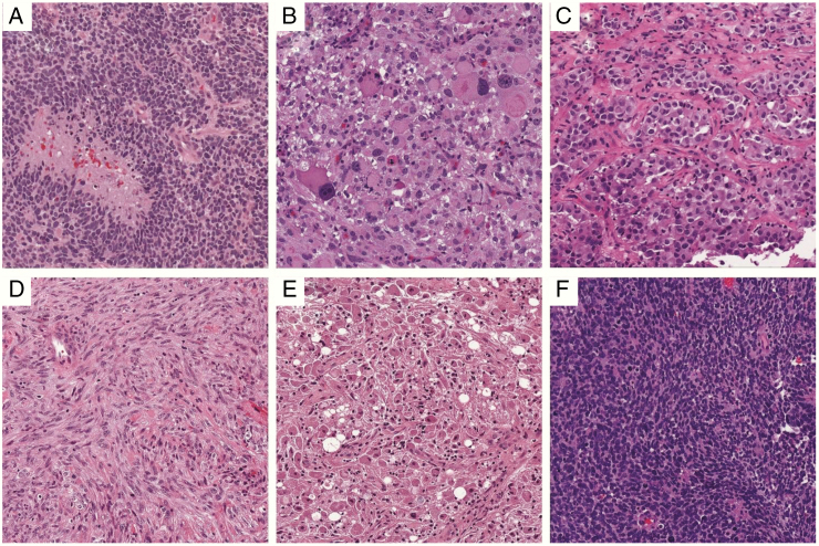
Fig. 3.
The many forms of GBM. (A) Classic GBM, with pseudopalisading necrosis and microvascular proliferation. (B) Giant cell GBM. (C) Epithelioid GBM with BRAF V600E. (D) Gliosarcoma. (E) Granular cell GBM. (F) Small cell GBM. All images are from the UPMC Neuropathology Virtual Slide Database, 200x magnification.

