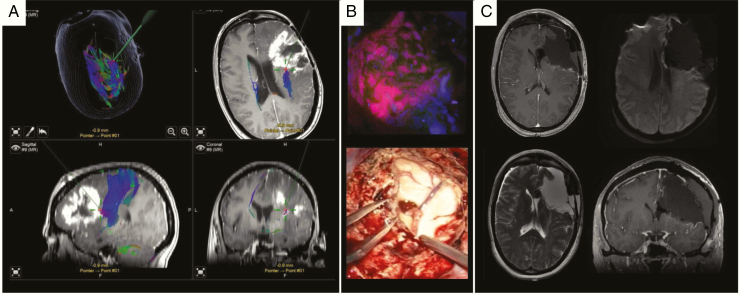
Fig. 8.
Microsurgical resection of a right-sided recurrent IDHwt glioblastoma WHO grade IV using intraoperative neuronavigation, neuromonitoring and 5-ALA fluorescence techniques. (A) T1 contrast enhanced axial, sagittal and coronal planes including DTI fiber tracking (blue fibers). The green trajectories/red points represent the pointer for intraoperative neuronavigation. (B) Upper image: corresponding intraoperative 5-ALA fluorescence image taken from the area as depicted by neuronavigation. Lower image: opening of the right ventricle due to critical involvement by tumor formation. (C) Postoperative MRI confirms gross total resection without residual contrast enhancement, no perilesional ischemia (diffusion-weighted image upper right).

