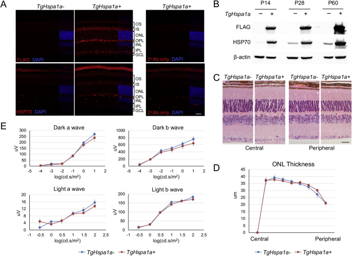
Figure 2.
Augmented expression of HSP70 from TgCrx-Hspa1a-Flag transgene in wild-type retina. (A) Immunofluorescent labeling of FLAG and HSP70 in retinal cross-sections from TgHspa1a+ mice and TgHspa1a- littermates at P28. (B) Immunoblots showing elevated expression of HSP70 protein driven by the TgCrx-Hspa1a-Flag transgene at P14, P28, and P60. (C) Representative picture of H&E stained retinal cross-sections, and (D) plot of ONL thickness measurements from TgHspa1a+ (n = 4) and TgHspa1a- littermates (n = 5) at 8 months. (E) ERG response in TgHspa1a+ (n = 3) and TgHspa1a- littermates (n = 6) at 8 months. Scale bar for all panels: 25 μm. Error bar: SEM. OS, outer segment; IS, inner segment; ONL, outer nuclear layer; OPL, outer plexiform layer; INL, inner nuclear layer; IPL, inner plexiform layer; GCL, ganglion cell layer.

