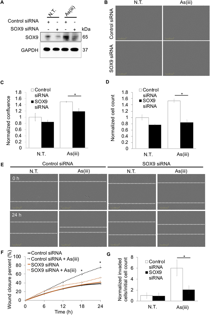
Figure 7: Loss of SOX9 negates the metastatic potential of arsenic-transformed lung epithelial cells.
BEAS-2B NRF2+/+ cells that were either untreated or received 0.5 μM arsenic (As(iii)) treatment for 30 passages (~3 months) were transiently transfected with either 40 nM control or 40 nM SOX9 siRNA for 72 hours. (A) Immunoblot analysis of SOX9 protein levels. (B) Representative images of cell confluence. (C) Quantification of images in (B) (n=3). (D) Cells were counted 24 hours post seeding (n=3). (E) Representative images of cells at 0 and 24 hours post scratch; cells were scratched at 72 hours post siRNA transfection (white dashed lines indicate scratch edges). (F) Wound closure percent was calculated at 0, 6, 12, 18, and 24 hours post scratch using same conditions at (E) (n=3). (G) Cells were serum starved overnight and at 72 hours post transfection were measured for invasion via number of cells that migrated into bottom chamber 24 hours later compared to initial starting number of cells (n=4–6); (*p<0.05).

