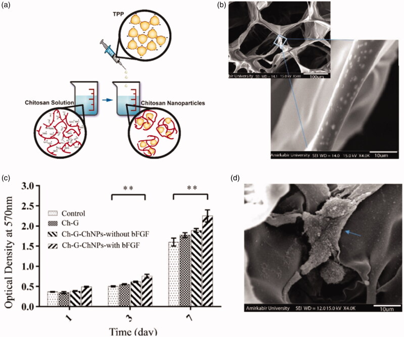
Figure 2.
(a) A schematic of chitosan nanoparticles prepared by ionic gelation method; (b) SEM showed the chitosan nanoparticles were incorporated within the chitosan-gelatin scaffold; (c) MTT assay for fibroblast cells cultured on scaffolds after 1, 3 and 7 days (**p < .01); (d) SEM image of fibroblast cells attached to the chitosan nanoparticles loading growth factors in chitosan-gelatin scaffold on day 7 after culture; The blue arrow stands for the attached cells (Azizian et al., 2018).

