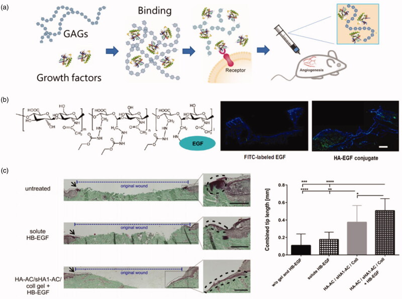
Figure 3.
(a) Growth factors binding with GAGs (Hachim et al., 2019). (b) the chemical structure of hyaluronate-epidermal growth factor (HA-EGF) conjugates; the fluorescence graphs of the degradation behavior of EGF and HA-EGF conjugate in HA films after topical application of FITC-labelled samples on wounded tissues (Niu et al., 2019). (c) HB-EGF released from sHA1-AC-containing hydrogels promotes epidermal tip formation and the results were measured by Masson-Goldner Trichrome staining. Bars: 500 μm; 100 μm (insets). The combined tip lengths of both wound sites were shown (n = 3 with 3–4 sections per condition *p < .05, **p < .01, ***p < .005, ****p < .001) (Thönes et al., 2019).

