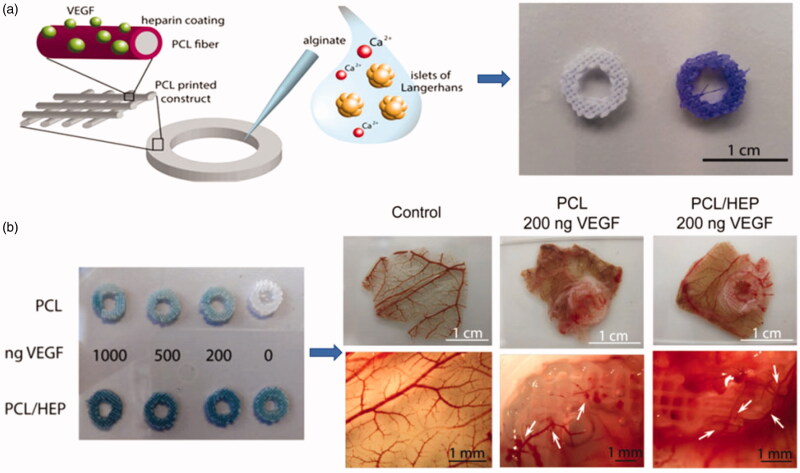
Figure 4.
(a) A schematic of showing the functionalized surface of PCL constructs binding VEGF. And the PCL and PCL/heparin plotted scaffolds stained with Azure II. (b) Graphs of neovascularization in PCL-VEGF and PCL-heparin-VEGF (Marchioli et al., 2016).

