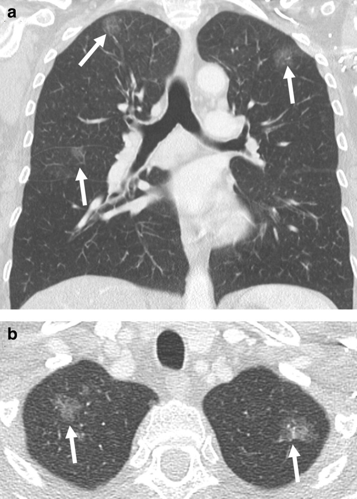
Figure 5.
(a) Coronal and (b) axial contrast-enhanced CT of a female patient with three pure ground-glass nodules that represent in situ adenocarcinomas in the right and left upper lobe, as well as the middle lobe (arrow). The correct staging is based on the tumour dimension of the largest ground-glass nodules (3.3 cm), as well as the number of tumours, which is given in brackets: T2a(3)N0M0.

