
Figure 9.
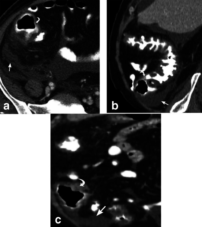
Pseudomembranous colitis. 54-year-old male presents with diarrhea for 3 days. Diffuse colonic wall thickening involving the cecum (curved white arrows) is depicted on axial CT (a) and sagittal MPR CT images (b, c). There is surrounding inflammatory soft tissue stranding and free fluid (straight white arrows). Subsequent stool assay reveals that the patient was positive for the C. difficile toxin. Sigmoid biopsy reveals colonic mucosa with superficial necrosis and fibrinopurulent exudate. MPR, multiplanar reconstruction.