
FIGURE 2.
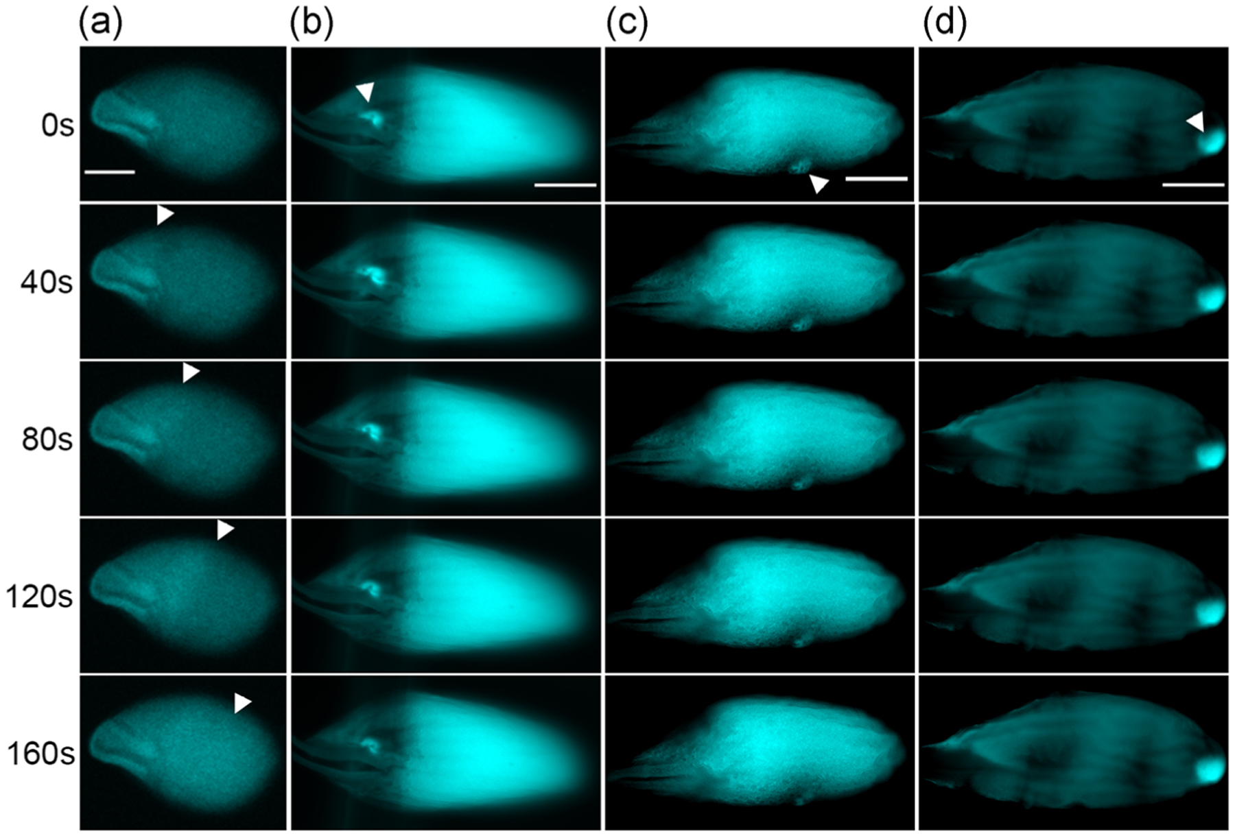
Regional calcium rise induced by microneedles in wildtype oocytes and calcium waves in kug mutant oocytes. (a) Representative images showing calcium waves propagating from the ends of kug mutant near-spherical shaped mature oocytes (n = 15). Arrowheads: wavefront of the calcium wave. (b–d) Representative images showing regional calcium rises that do not propagate into waves induced by microneedle pressing at (b) anterior (n = 7), (c) waist (n = 4), and (d) posterior (n = 4) regions of wildtype mature oocytes. Arrowheads: pressing site of microneedles. The background level of calcium varies among mature oocytes (Hu & Wolfner, 2019); scale bar = 100 μm