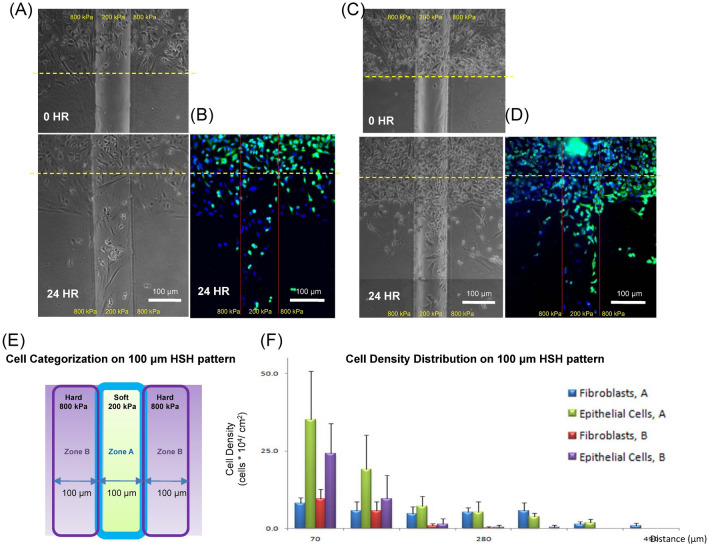
Figure 7.
A549 epithelial cells and WI-38 fibroblasts co-cultured motility for the vertical configuration with cells moving along the length of the rectangular strip with a width of 100 μm in our HSH system. (A) The co-culture was imaged with phase contrast at 0 h and 48 h after being released. (B) Immunofluorescent images were captured after 24 h as well. A second representative response of the A549 epithelial cells and WI-38 fibroblast response at (C) 0 and 48 h, and (D) with immunofluorescence. (E) A diagram indicating the location of zone A and zone B on the HSH substrate for (F) quantifying the motility of the A549 epithelial cells and WI-38 fibroblasts on the vertical HSH substrate. Green pseudo-coloring for was for Cytokeratin and A549 epithelial cells and blue pseudo-coloring was for DAPI and the nucleus. The differences between the two cell types were statistically significant (P value = 10–24) for the cells located inside the 100 μm wide soft strip (group A).

