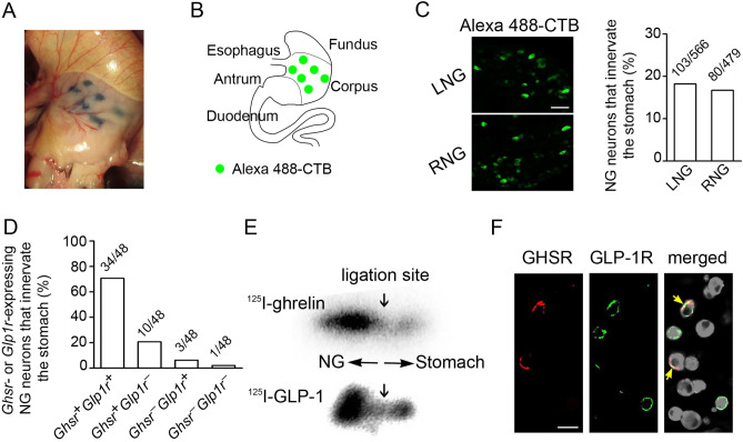
Figure 2.
Transportation of GHSR and GLP-1R through the vagal afferent nerve in mice. (A) Representative photo to demonstrate multiple injection sites of Alexa Fluor 488-conjugated CTB in blue-black color at the gastric corpus. (B) Schematic illustration of injection sites of Alexa 488-CTB in green color at the ventral gastric corpus. (C) Representative images of left (LNG) and right (RNG) NG neurons labeled with Alexa Fluor 488-CTB transported from the gastric corpus. Percentage of stomach-projecting NG neurons (LNG; n = 566, RNG; n = 479). (D) Percentage of Ghsr- or Glp1r-expressing NG neurons expressing Ghsr or Glp1r that innervate the stomach (n = 48). (E) Representative autoradiographs showing the bindings of 125I-ghrelin and 125I-GLP-1 on the NG side of the ligated vagal afferents. (F) Representative images of immunocytochemistry of GHSR (red), GLP-1R (green), and their colocalization (arrows in yellow) in dispersed NG neurons. Scale bars 50 µm (C) and 25 µm (F).

