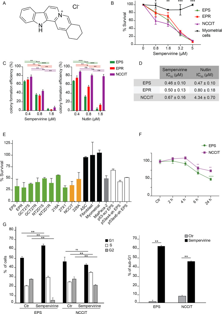
Fig. 1. Sempervirine induces cell death in p53-wt and p53-null germ cell tumor lines.
A Chemical structure of sempervirine chloride. B Mean half-maximal inhibition of cell survival/proliferation (IC50) of sempervirine after 72 h of culture in 2102EP(S) (EPS), 2102EP(R) (EPR), NCCIT, and myometral cells. *p < 0.05, **p < 0.01, ***p < 0.001, unpaired t test, number of biological replicates (n) n = 5. C Colony formation assay on 2102EP(S) (EPS), 2102EP(R) (EPR), and NCCIT after treatment with 0.4, 0.8, and 1.6 μM of sempervirine and nutlin, respectively. ****p < 0.0001, ***p = 0.0006. Two-way ANOVA, Tukey’s multiple comparisons test, n = 3. D IC50 of sempervirine and nutlin treatment in 2102EP(S) (EPS), 2102EP(R) (EPR), and NCCIT. E Proliferation assay on several TGCT cell lines p53-wt (green) and p53-mut or -null (yellow), MiaPaCa-2 (gray), control cell lines (black), and silenced 2102EP(S) (white) treated with 5 μM sempervirine for 24 h. Mean ± SD, n = 3. F Reversibility after 2, 4, 6, or 24 h of treatment with 5 μM sempervirine. **p < 0.01, ****p < 0.0001, paired t test, n = 3. G FACS analysis of sempervirine-treated 2102EP(S), and NCCIT cells showing the percentage of cells in G1, S, G2 cell cycle phases (left) and the percentage of sub-G1 cells (right) after 72 h of treatment. *p < 0.05, **p < 0.01, unpaired t test, n = 3.

