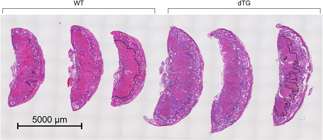
Fig. 3: Placenta histology from WT and dTG mice,
The images show 3 placentas from a WT litter juxtaposed to 3 placentas from a dTG litter, obtained at E17.5 and stained with hematoxylin and eosin. The border between the spongiotrophoblast and the labyrinth is outlined by a dashed black line in one WT placenta and one dTG placenta.

