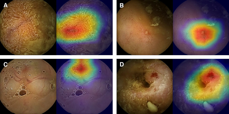
Fig 3. Concordance of significant images between the AI and experts.
All the four images were classified as significant with 0.8 or higher probability and based on manual classification by experts (left side of the images). As the color spectrum of class activation map turns red, lesions display higher probabilities. AI can distinguish multiple findings that coexist in an image (right side of the images; A, swollen villi from debris; B, Small mucosal defect from the nearby debris; C, Vascular tuft adjacent to vessels; D, Vascular tuft surrounded by inflamed mucosa).

