
Figure 4.
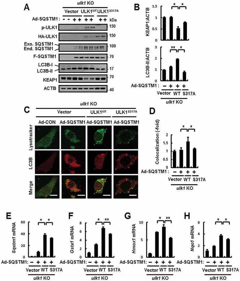
SQSTM1-mediated ULK1 phosphorylation induces autophagy activation. (A) ulk1 KO MEFs co-transfected with vectors encoding HA-WT ULK1 or the HA-ULK1S317A mutant were infected with Ad-SQSTM1 for 18 h and subjected to immunoblot analysis with antibodies against p-ULK1(S317), HA-ULK1, SQSTM1, FLAG-SQSTM1, LC3B, KEAP1, and ACTB (loading control). (B) Densitometric analysis of KEAP1 and LC3B-II immunoblots. (C) Confocal microscopy analysis of colocalization of Lysotracker and LC3B the cells treated as described in (A). The representative single optical sections and merge images are shown. Scale bar: 10 μm. (D) Quantitative analysis of colocalization. (E–H) Total mRNA isolation from cells treated as described in (a) and subjected to qRT-PCR analysis for relative mRNA expression of sqstm1 (E), Gsta1 (F), Hmox1 (G), and Nqo1 (H). Data are presented as the mean ± SD from 3 independent experiments. *p < 0.05, **p < 0.01